
Have you ever asked yourself, “What is IQ?” Historically, IQ (intelligence quotient) was defined as a score or number that is calculated by dividing your mental age by your chronological age and multiplying it by 100. If that did not clarify things or you felt that was a not satisfactory answer to my original question, perhaps the following definition may help. Dr. David Wechsler, a clinical psychologist who developed one of the most well-known IQ tests (Wechsler Scales of Intelligence) defined intelligence as an individual’s ability to adapt to and constructively solve problems in the environment. Specifically, he stated that intelligence was “the aggregate or global capacity of the individual to act purposefully, to think rationally, and to deal effectively with his environment” (Wechsler, 1939). In essence, he portrayed intelligence in terms of performance rather than capacity or quantity. He felt it was more important how well one uses his/her intelligence compared with how much intelligence you have.
Another intriguing question is, “Where is IQ located in the brain?” Recently, researchers have attempted to answer this question using brain-imaging techniques. At the University of Illinois, neuroscience researcher Dr. Aron Barbey published results from a study examining the physical architecture of intelligence and the brain (Brain, 2012). He attempted to identify brain structures that are vital to general intelligence and aspects of intellectual functioning. His method of approach was to study individuals with focal brain injuries (e.g., Vietnam veterans with penetrating head injuries). Dr. Barbey felt that studying how damaged brain regions produce specific forms of cognitive impairment would allow him to map the architecture of the mind by identifying brain structures that are important critically for specific intellectual abilities. As shown in the figure below, Dr. Barbey found brain structures on the left side to be vital for IQ. One critique of this study is that utilizing research subjects with brain damage could skew test results, particularly if there is recovery of function utilizing other brain structures to compensate for the original damage.
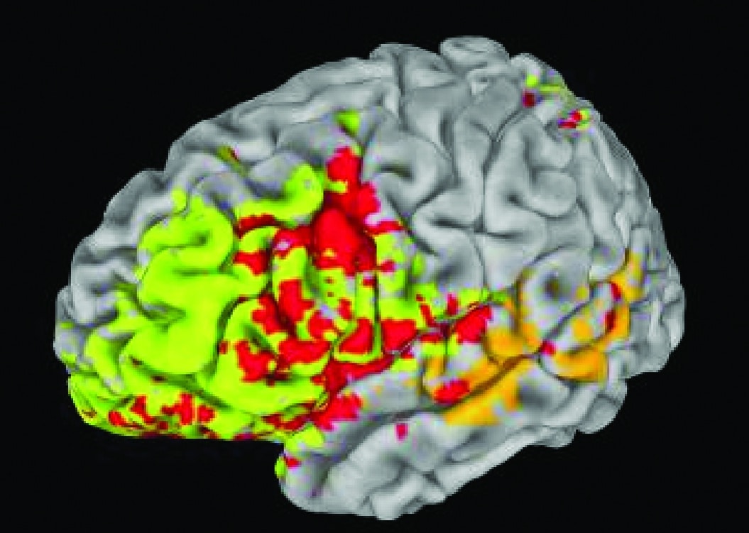
Figure 1. Dr. Barbey found brain structures on the left side to be vital for general intelligence (IQ) and executive function (EF). Red = common areas for IQ and EF; orange = IQ; yellow/green = EF.
In another study published in 2010 (Proceedings of the National Academy of Sciences), a research team from CalTech, USC and the University of Iowa also examined brain-lesion patients finding that IQ is determined by a network of regions across both sides of the brain rather than residing in a single structure within the brain. As shown in the figure below, these networks reach across the upper and lower sections of the brain. Again, this research utilized patients with pre-existing brain disorders, so it is not completely clear how these results apply to typical adults.
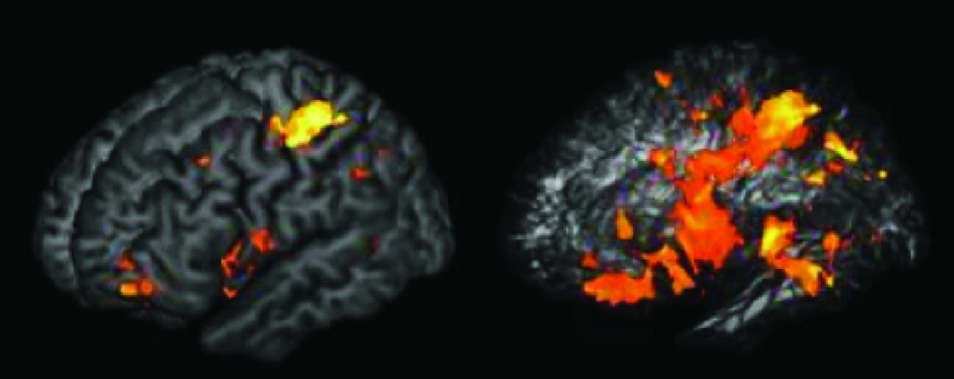
Figure 2. Brain regions important for IQ. The big orange regions in the right image (transparent brain) are connections (like cables) that link the specific brain regions in the image on the left (Gläscher et al, 2012).
In 2009, neuroscientist Paul Thompson from UCLA examined pairs of fraternal and identical twins without brain injury or impairment and found the layer of insulation coating neural wiring in the brain plays a critical role in determining IQ (Journal of Neuroscience). The layer of insulation is called myelin; think of it as the insulation on an electrical wire that keeps current from leaking out and boosts speed. Utilizing a new brain scan called diffusion-tensor imaging (DTI), these researchers found a strong correlation between the integrity of the myelin or white matter in the brain and performance on IQ tests (see figures below).
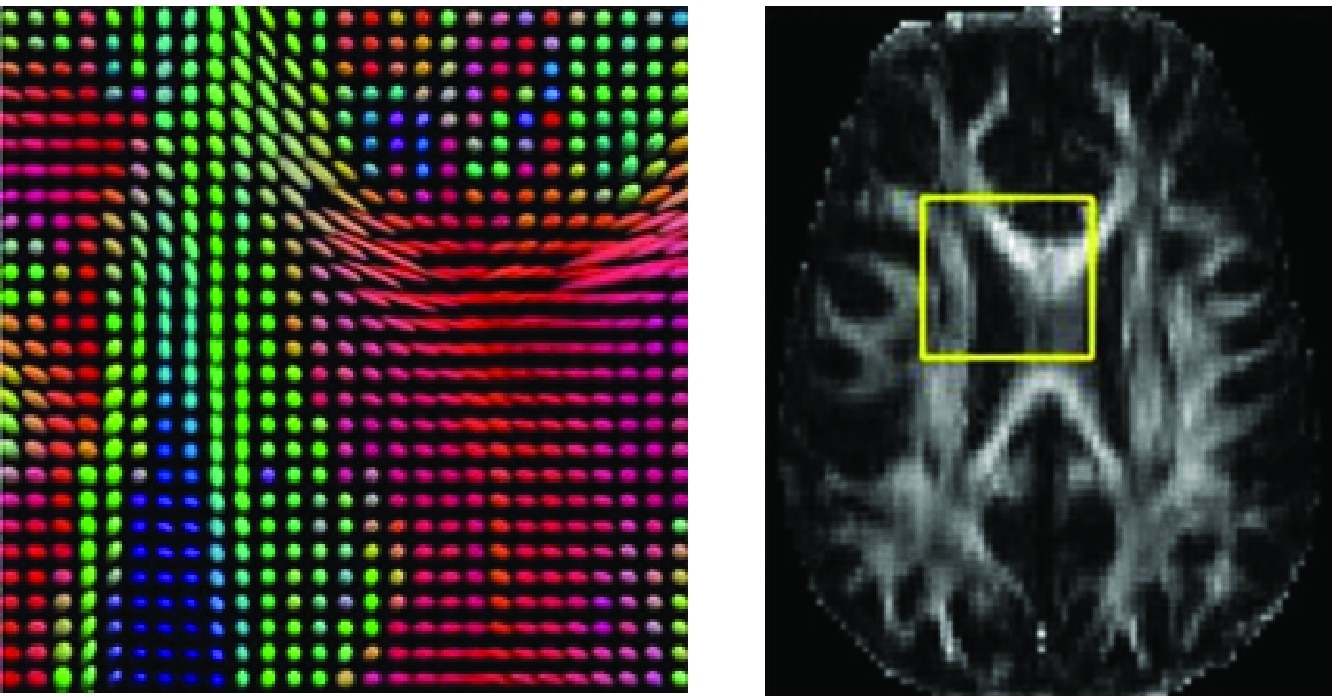
Figure 3. Pixilated brain: At the right, an MRI image shows a slice of the human brain. At the left, a magnified portion of this section is shown, created using diffusion imaging. To create the image, scientists measured the direction of the water diffusion in brain tissue. The “flower petals” at each point show the directions of fastest diffusion. These are aligned along the neural pathways of the brain, because water diffuses directionally along the well-insulated neural wires that carry electrical signals. The different directions of diffusion are color-coded red, green, and blue. In this example, the bright red areas reveal the thick fiber tract, called the corpus callosum, which transfers information between the left and right sides of the brain.
More recently, Dr. Angela Duckworth of the University of Pennsylvania has explored the impact of motivation on how well people perform on IQ tests. She found that competencies other than IQ predict academic and professional success, particularly the person’s desire to succeed (2013). Her conclusion was that how hard people try could be just as important to success in life as IQ. She coined the term “grit” to describe one’s ability to withstand stress and move past failure to achieve a goal. In her mind, grit is the best indicator of future success, not IQ. Her conclusions fit nicely with Dr. Wechsler’s viewpoint that it is not how much IQ you have, but how you use it (perseverance), that counts.

image from J. Irby
